


Oxytocin (2mg)
$25.00
Out of stock
Join Waitlist
Enter your email address to be notified when this item is back in stock.
OXYT-2
Description
Chemical Information
Shipping Information
What is Oxytocin?
Oxytocin is a naturally occurring nonapeptide hormone composed of nine amino acids with the molecular structure Cys-Tyr-Ile-Gln-Asn-Cys-Pro-Leu-Gly-NH₂. It is synthesized in the hypothalamus and released into the bloodstream by the posterior pituitary gland in mammals, where it plays a central role in physiological and behavioral functions.
In research settings, synthetic oxytocin is commonly used to study pathways related to social behavior, emotional regulation, stress response, and reproductive physiology.
As a research compound, oxytocin is prized for its well-characterized structure, high solubility in aqueous solutions, and stability when stored under appropriate conditions. Its activity is mediated via the oxytocin receptor (OXTR), a G-protein coupled receptor expressed in the brain, uterus, heart, and other tissues.
These properties make oxytocin an attractive model peptide for studying both central nervous system and peripheral signaling pathways.
Disclaimer: This oxytocin peptide product is intended for laboratory research purposes only. It is not approved for human or veterinary use. |
Mechanism of Action (Based on Research)
Oxytocin exerts its effects primarily by binding to the oxytocin receptor (OXTR), a member of the G-protein coupled receptor (GPCR) family. OXTR is expressed in various tissues, including the uterus, mammary glands, heart, and brain.
Upon binding, the receptor activates Gq proteins, which in turn stimulate phospholipase C (PLC). This enzyme catalyzes the conversion of PIP₂ into IP₃ and DAG, leading to an increase in intracellular calcium levels and activation of protein kinase C (PKC).
In smooth muscle tissues, such as the myometrium (uterine muscle), this calcium surge initiates muscle contraction — a critical process during labor. In mammary glands, oxytocin induces contraction of myoepithelial cells to promote milk ejection.
Across systems, oxytocin’s activity is dose-dependent, tissue-specific, and influenced by receptor density, hormonal state, and route of administration. This complex MoA makes oxytocin a versatile peptide for research exploring both endocrine and neurobehavioral functions.
Effects on the CNS
In the central nervous system, oxytocin acts on neurons in regions like the amygdala, hypothalamus, and prefrontal cortex.
Although the precise downstream pathways are still being studied, oxytocin appears to modulate neuronal excitability and synaptic transmission, which in turn influences social bonding, emotional processing, trust behavior, and stress regulation.
Oxytocin has been observed to modulate social cognition, trust, pair bonding, and stress reactivity. Research using rodent and primate models shows that oxytocin enhances pro-social behavior, likely by influencing neural circuits in the amygdala, prefrontal cortex, and hypothalamus [1].
In primates, oxytocin has also been demonstrated to enhance social attention and reinforcement in rhesus macaques, involving amygdala‑prefrontal circuits [2].
Action on Smooth Muscles
In peripheral systems, oxytocin plays key roles in uterine contraction during labor and milk ejection during lactation, acting on receptors in uterine smooth muscle and mammary glands [3].
Oxytocin also modulates cardiovascular tone, wound healing, and anti-inflammatory signaling, though these effects are still under investigation in preclinical studies [4].
Research Applications of Oxytocin (Preclinical Observations)
Although oxytocin has not been approved for therapeutic use, extensive non-clinical studies highlight its observed effects across multiple research models.
These findings offer insights into its potential utility in neuroscience, inflammation modulation, cardiovascular protection, and tissue repair.
Social Behavior & Neuroprotection
Preclinical investigations in rodents and primates have consistently demonstrated oxytocin’s role in social cognition and bonding.
In prairie voles, limbic oxytocin release during pair-bonding facilitates long-term partner affinity; analogous pathways are implicated in trust and empathy models in humans and dogs, often studied using intranasal or central oxytocin administration [5].
Emerging research also suggests oxytocin may protect the brain in stroke and ischemia models [6]. In socially enriched rodents, oxytocin signaling attenuates infarct size and reduces markers of neuronal inflammation and oxidative stress, mediated via microglial modulation [7].
Anti‑Inflammatory & Immune Modulation
Animal models indicate that oxytocin exerts measurable anti-inflammatory effects across various tissues.
In murine obesity models, chronic oxytocin infusion reduced visceral macrophage infiltration, adipocyte hypertrophy, and expression of pro‑inflammatory cytokines (e.g., IL‑6, TNF‑α), while boosting adiponectin levels in the serum [8].
Oxytocin also restrained acute inflammatory responses in models of localized inflammation. In a rat air-pouch model, oxytocin significantly decreased exudate volume, leukocyte infiltration, and VEGF and IL‑1β levels—effects comparable to standard NSAID treatment [9].
In cellular models, oxytocin attenuated microglial activation and cytokine release (e.g. IL‑6, TNF‑α) via ERK/p38 MAPK signaling pathways, suggesting a potential role in dampening neuroinflammatory processes [7].
Cardiovascular & Metabolic Regulation
Oxytocin has been linked to cardioprotection in models of myocardial ischemia-reperfusion injury.
Studies report improved post-infarct ventricular function, reduced fibrosis and cell death, enhanced capillary density, and activation of cardiomyocyte survival pathways via PI3K/Akt signaling. These effects appear mediated through nitric oxide and atrial natriuretic peptide pathways [4][8].
Alongside cardiac effects, oxytocin infusion in dyslipidemic and obese rodent models has shown reduced progression of atherosclerosis, lower systemic CRP levels, and restricted adipose tissue inflammation—implicating oxytocin signaling in broader vascular and metabolic regulation [7][9].
Tissue Repair & Wound Healing
Various preclinical studies observe oxytocin’s involvement in tissue repair and regeneration. Reports include improved healing in wound, flap, and gastric injury models and accelerated epithelial recovery across multiple organs.
Some of these effects are thought to involve vagal signaling induced by microbial metabolites (e.g. L. reuteri), highlighting an interplay between microbiota and oxytocin-mediated tissue homeostasis [10].
Oxytocin Peptide Characteristics
- Molecular Formula: C₄₃H₆₆N₁₂O₁₂S₂
- CAS Number: 50‑56‑6
- Amino Acid Sequence: Cys–Tyr–Ile–Gln–Asn–Cys–Pro–Leu–Gly–NH₂ (cyclic nonapeptide with disulfide bridge between Cys¹ and Cys⁶)
- Synonyms: OXT, OT, Oxytocin Acetate, Neuropeptide Oxytocin
- Molar Mass: ~1007.2 g/mol
- Form: Lyophilized peptide powder
- Recommended Storage: –4°F (–20 °C) for long-term; 35.6–46.4°F (2–8 °C) for short-term (avoid repeated freeze–thaw cycles)
Oxytocin vs Arginine Vasopressin vs LIT-001 Comparison
Feature | Arginine Vasopressin (AVP) | LIT‑001 | |
Type | Endogenous nonapeptide hormone (OXT receptor agonist) | Endogenous nonapeptide hormone (V₁A/V₂ receptor agonist) | Synthetic small-molecule OXTR agonist |
Primary Targets | Oxytocin receptor (OXT‑R); partly V₁A at high doses | Vasopressin V₁A/V₁B/V₂ receptors; minor cross‑reactivity | Oxytocin receptor selectively (agonist) |
Mechanism Complexity | Single-receptor GPCR (OXT‑R), calcium signaling cascade | Multi-receptor GPCR family (V₁A, V₂, V₁B); complex access | Single-receptor agonist; improved CNS penetration |
Research Focus | Social behavior, neuroendocrine regulation, uterine/milk effects | Fluid balance, vasoconstriction, social/emotional behavior | CNS-based social function models |
Pharmaco- kinetics | Rapid clearance (20–60 min half‑life), poor oral absorption | Peptide; similar limitations to oxytocin | Enhanced CNS exposure versus OXT; brain penetrance (~25 nM EC₅₀) |
Research Stage | Extensively used in preclinical & some clinical trials | Widely studied in physiology and behavior; some clinical use | Preclinical; early-stage behavioral pharmacology models |
Additional Observed Effects | Social bonding, stress modulation, uterine contraction, lactation | Fluid homeostasis, circadian modulation, social stress, blood pressure regulation | Mimics oxytocin social behavior effects; better receptor specificity |
Regulatory Status | Research use only | Endogenous hormone; lab reagent; therapeutic analogues exist | Research use only |
Disclaimer | Not for human/veterinary use | Not for human/veterinary use unless medical-grade formulation | Research tool only; not for human consumption |
Oxytocin Peptide Safety (Research Settings)
In non-clinical animal models, oxytocin has demonstrated a relatively favorable safety profile at research-level dosages. Rodent studies, for example, have noted minimal adverse effects when oxytocin is administered intranasally or subcutaneously in controlled environments [11].
Common endpoints evaluated include behavior, reproductive tissue activity, and cardiovascular markers. Most observed effects have been dose-dependent and transient, with no indication of long-term toxicity at moderate exposure levels in short-term trials.
However, in preclinical and clinical settings, high-dose or prolonged oxytocin administration beyond physiological levels has been associated with electrolyte disturbances—notably hyponatremia and water intoxication—via natriuretic mechanisms in rodent models.
These effects reflect oxytocin’s antidiuretic-like activity and can manifest alongside hemodynamic instability in some obstetric administration cases. Additionally, excessive oxytocin has been linked to uterine hyperstimulation, tetanic contractions, and uterine rupture in obstetric contexts.
Early rodent and primate studies also suggest that prolonged exposure may blunt behavioral responsiveness [12].
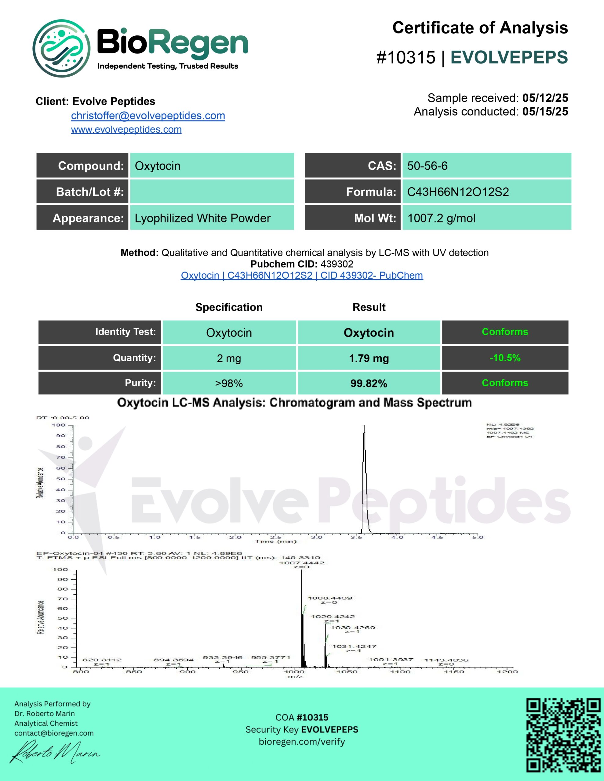
Certificate of Analysis (COA)
Each vial of Oxytocin Peptide is backed by a Certificate of Analysis (COA) issued by a qualified third-party laboratory. A COA verifies:
- Peptide identity and molecular integrity
- Purity levels (typically ≥99%)
- Peptide concentration per vial
- Screening results for contaminants such as heavy metals, microbial agents, and endotoxins
Evolve Peptides makes every COA available to customers—either as a downloadable PDF directly from the product page or upon request via support. Testing is conducted in ISO/GLP-certified labs using validated analytical methods, including HPLC and mass spectrometry, to ensure reproducible quality from batch to batch.
Legal Disclaimer
Oxytocin peptide is intended solely for laboratory research use only. It is not approved for human or veterinary use. Is it not for resale, diagnostic, therapeutic, or clinical applications.
By purchasing this product, you acknowledge and assumes full responsibility for
the safe handling, storage, and disposal of this material. Use of this product in violation of any local, state, or federal laws is strictly prohibited.
Scientific References
- Hiroyuki Arakawa, Dynamic regulation of oxytocin neuronal circuits in the sequential processes of prosocial behavior in rodent models, Current Research in Neurobiology, Volume 2, 2021, 100011, ISSN 2665-945X, https://www.sciencedirect.com/science/article/pii/S2665945X21000073)
- Gangopadhyay P, Chawla M, Dal Monte O, Chang SWC. Prefrontal-amygdala circuits in social decision-making. Nat Neurosci. 2021 Jan;24(1):5-18. Epub 2020 Nov 9. PMID: 33169032; PMCID: PMC7899743. https://pmc.ncbi.nlm.nih.gov/articles/PMC7899743/
- Walter MH, Abele H, Plappert CF. The Role of Oxytocin and the Effect of Stress During Childbirth: Neurobiological Basics and Implications for Mother and Child. Front Endocrinol (Lausanne). 2021 Oct 27;12:742236. PMID: 34777247; PMCID: PMC8578887. https://pmc.ncbi.nlm.nih.gov/articles/PMC8578887/
- Jankowski M, Broderick TL, Gutkowska J. The Role of Oxytocin in Cardiovascular Protection. Front Psychol. 2020 Aug 25;11:2139. PMID: 32982875; PMCID: PMC7477297. https://pmc.ncbi.nlm.nih.gov/articles/PMC7477297/
- Blumenthal SA, Young LJ. The Neurobiology of Love and Pair Bonding from Human and Animal Perspectives. Biology (Basel). 2023 Jun 12;12(6):844. PMID: 37372130; PMCID: PMC10295201. https://pmc.ncbi.nlm.nih.gov/articles/PMC10295201/
- Momenabadi S, Vafaei AA, Zahedi Khorasani M, Vakili A. Pre-Ischemic Oxytocin Treatment Alleviated Neuronal Injury via Suppressing NF-κB, MMP-9, and Apoptosis Regulator Proteins in A Mice Model of Stroke. Cell J. 2022 Jun;24(6):337-345. https://pmc.ncbi.nlm.nih.gov/articles/PMC9315214/
- Yuan, L., Liu, S., Bai, X. et al. Oxytocin inhibits lipopolysaccharide-induced inflammation in microglial cells and attenuates microglial activation in lipopolysaccharide-treated mice. J Neuroinflammation 13, 77 (2016). https://jneuroinflammation.biomedcentral.com/articles/10.1186/s12974-016-0541-7
- Szeto, A., Cecati, M., Ahmed, R. et al. Oxytocin reduces adipose tissue inflammation in obese mice. Lipids Health Dis 19, 188 (2020). https://lipidworld.biomedcentral.com/articles/10.1186/s12944-020-01364-x
- Jankowski M, Bissonauth V, Gao L, Gangal M, Wang D, Danalache B, Wang Y, Stoyanova E, Cloutier G, Blaise G, Gutkowska J. Anti-inflammatory effect of oxytocin in rat myocardial infarction. Basic Res Cardiol. 2010 Mar;105(2):205-18. https://pubmed.ncbi.nlm.nih.gov/20012748/
- Brown, C. H. (2025). Is oxytocin “nature’s medicine”? Pharmacological Reviews. Advance online publication. https://pharmrev.aspetjournals.org/article/S0031-6997%2824%2900978-5/fulltext
- Disala Fernando, Sarah Siederer, Sunita Singh, Ian Schneider, Ashutosh Gupta, Marcy Powell, Duncan Richards, Michelle P. McIntosh, Peter Lambert, Susan Fowles, Safety, Tolerability and Pharmacokinetics of Single Doses of Oxytocin Administered via an Inhaled Route in Healthy Females: Randomized, Single-blind, Phase 1 Study, EBioMedicine, Volume 22, 2017, Pages 249-255, ISSN 2352-3964. https://www.sciencedirect.com/science/article/pii/S2352396417302931
- Gangopadhyay P, Chawla M, Dal Monte O, Chang SWC. Prefrontal-amygdala circuits in social decision-making. Nat Neurosci. 2021 Jan;24(1):5-18. https://pmc.ncbi.nlm.nih.gov/articles/PMC7899743/ https://pubmed.ncbi.nlm.nih.gov/31393999/
Contents: 2 mg lyophilized (freeze-dried) powder provided in a 3 ml vial, sealed and sterile. Purity exceeds 99%, guaranteed.
Notes:Requires reconstitution with bacteriostatic water. (Sold Here:BAC Water.)
Chemical Formula: C78H123N23O23S2
PubChem CID: 71300630
CAS Number: 386264-39-7
Molecular Weight: 1815.12 g/mol
Storage:Store at 8C, sealed, away from heat, light, and moisture. The colder the better.
Purity:>99%
Orders placed before 1 PM EST ship same day.
We use USPS for most of our deliveries. You can expect your parcel within 2-5 business days from when it leaves our warehouse.
Why Choose Us?
Lyophilized in Texas – 99%+ Purity Guaranteed
Our peptides are proudly lyophilized right here in the US, to exact specifications. Strictly no compromises.
Rigorously Third-Party Tested, Always
Every product page includes independent lab test results for every peptide we sell.
We Respond in Minutes, Not Days
We take pride in providing excellent support – we're here for your total research success.
200% Money-Back Guarantee
Test our product – if purity doesn't match the label, we refund 200%. No questions asked.
Related products
-

-

 Save $45.00
 -

 Save $25.00
BPC-157 10 mg
$79.90Original price was: $79.90.$54.90Current price is: $54.90.Save $25.00 -